
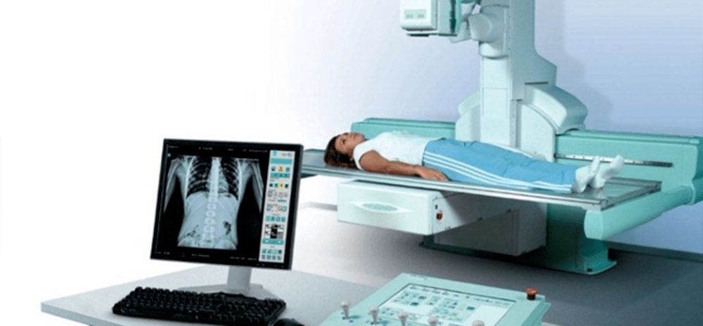
Fluoroscopy is a medical imaging technique that uses X-rays to obtain real-time moving images of the internal structures of a patient through the use of a fluoroscope. In the past, fluoroscopy systems used image intensifier technology and analog video to display and record these moving X-ray images. However, over the past decade digital fluoroscopy systems have begun replacing their analog predecessors. These new digital systems provide many advancements in image quality, dose efficiency, and workflow compared to traditional analog fluoroscopy.
Advancements in Image Quality
One of the main benefits of digital fluoroscopy systems is the improvements they offer in image quality compared to analog systems. Digital fluoroscopy uses a flat panel digital detector instead of an image intensifier, directly converting the X-ray photons into a digital image. This results in higher resolution, sharper images with less noise and artifacts. Digital detectors have a higher detective quantum efficiency, which means they are able to detect more of the X-ray information that reaches the detector. Luminescent materials in the detector convert X-ray energy directly into visible light photons which are then measured by tiny sensor elements. This allows the full resolution capabilities of the detector to be utilized rather than being limited by analog video processing like in older systems. Additionally, digital imaging technologies like dose grids can be used to further improve low contrast resolution without increasing patient dose. The high-definition images produced by digital fluoroscopy benefit clinicians in diagnosis and guidance during interventional procedures.
Increased Dose Efficiency
Another critical advantage of Digital Fluoroscopy System is their improved dose efficiency compared to analog systems. Digital detectors have a wider dynamic range, which allows them to achieve proper exposures with lower X-ray doses. Features like automatic brightness control and last image hold can adjust exposure factors pulse by pulse based on prior images, optimizing dose delivery. Digital systems also support leading dose reduction technologies like asymmetric collimation that restrict the X-ray field only to the clinical area of interest. Imaging hardware and software work together utilizing factors like automatic kV selection and variable focal spot size depending on patient size and clinical needs. Depending on the examination and patient, estimates show digital fluoroscopy can reduce radiation exposure to the patient by 30-50% compared to equivalent exams on older analog systems. The lower doses are an important consideration especially for pediatric patients and long interventional procedures.
Streamlined Workflow and Archiving
Digital imaging has enabled faster, more streamlined workflows in clinical and interventional fluoroscopy procedures. Large, high-resolution monitors allow multi-modality review of all relevant imaging during a case without switching tapes or films. Clinicians have simultaneous access to fluoroscopy, roadmaps, and other previous exams. Digital images are immediately available for recording procedural videos, acquiring still frames, performing post-processing techniques like tomosynthesis, and performing measurements. Images and video clips can be wirelessly sent to other departments, shared securely with referring physicians for consultations, or stored permanently in electronic archives. Transitioning from physical films to digital archiving also improves space utilization and eliminates film reproduction or storage costs over the long term. DICOMnetworking capabilities support teleradiology and sharing of images and reports across the entire healthcare enterprise.
Application of Advanced Visualization Techniques
As digital technologies continue advancing, new visualization techniques are being introduced in Digital Fluoroscopy Systems to provide clinicians additional useful tools. Some applications include dual energy subtraction to filter out bone or soft tissue, tomosynthesis to acquire multiple low dose images through different planes and reconstruct serial slices, and three dimensional model creation from biplane systems. These add functionalities like virtual intraoperative navigation during complex procedures. Advanced quantification packages can also analyze fluoroscopy videos to track catheter or stent movement, dynamically measure vessel diameter changes, or calculate physiological parameters. The highly programmable digital platforms support development of customized applications tailored for specific clinical needs. As visualization becomes more sophisticated, it enhances clinicians’ capabilities to diagnose problems and guide interventions using live X-ray guidance.
Mobile C-Arms and Miniature Systems
Digital detectors have enabled the development of miniaturized mobile C-arms as well as smaller portable fluoroscopy systems. Wheel-mounted mini C-arms weigh under 55 pounds, facilitating their movement between operating rooms and saving staff effort. They support a variety of general, orthopedic, and trauma exams without moving the patient. Portable fluoroscopes are so compact that a single technician can transport them to the bedside, ICU, or emergency department. Both feature high-resolution flat panel detectors in a fraction of the size and weight of traditional C-arms. Digital imaging has removed previous constraints on miniaturization. Lower power consumption also allows extended battery life for fully wireless exams away from wall outlets. The mobility, flexibility and access provided by these new generation systems improve workflow and patient throughput in busy clinical settings. They expand the appropriate uses of fluoroscopy to areas traditionally relying only on non-interventional imaging.
Regulatory Changes Facilitating New Technologies
As digital technologies matured, revisions were needed to existing regulations which were mostly developed for analog fluoroscopy. Newer drafts of governing documents recognized digital imaging capabilities exceeding analog benchmarks. They incorporated recommendations validating the efficacy of advanced dose reduction and safety features found only in digital systems. Appropriate adjustments ensured regulations supported adoption of these new medical technologies rather than created barriers. Provided manufacturers comply with appropriate testing and performance verification standards, revised guidelines accommodate solutions like large flat panel detectors, higher resolution biplane systems, and new visualization software packages. Regulatory bodies worked closely with industry to balance patient safety with allowing progressive clinical capabilities. This collaborative approach established standardized evaluation methods, streamlining introduction of digital innovations and benefiting patient care.
Digital fluoroscopy systems have evolved rapidly over the past decade from their analog predecessors. The migration to digital technologies has significantly enhanced clinical capabilities through high resolution imaging, reduced radiation doses, more efficient workflows, versatile portability, and advanced visualization applications. However, there are still additional frontiers to explore as detector resolutions continue multiplying and computing power drives ever more sophisticated software packages. Close cooperation between manufacturers, clinicians, and regulators will remain important to ensure patients benefit from responsible progress towards the next generation of fluoroscopy technology.
*Note:
1. Source: Coherent Market Insights, Public sources, Desk research
2. We have leveraged AI tools to mine information and compile it

About Author – Vaagisha Singh
Vaagisha brings over three years of expertise as a content editor in the market research domain. Originally a creative writer, she discovered her passion for editing, combining her flair for writing with a meticulous eye for detail. Her ability to craft and refine compelling content makes her an invaluable asset in delivering polished and engaging write-ups. LinkedIn