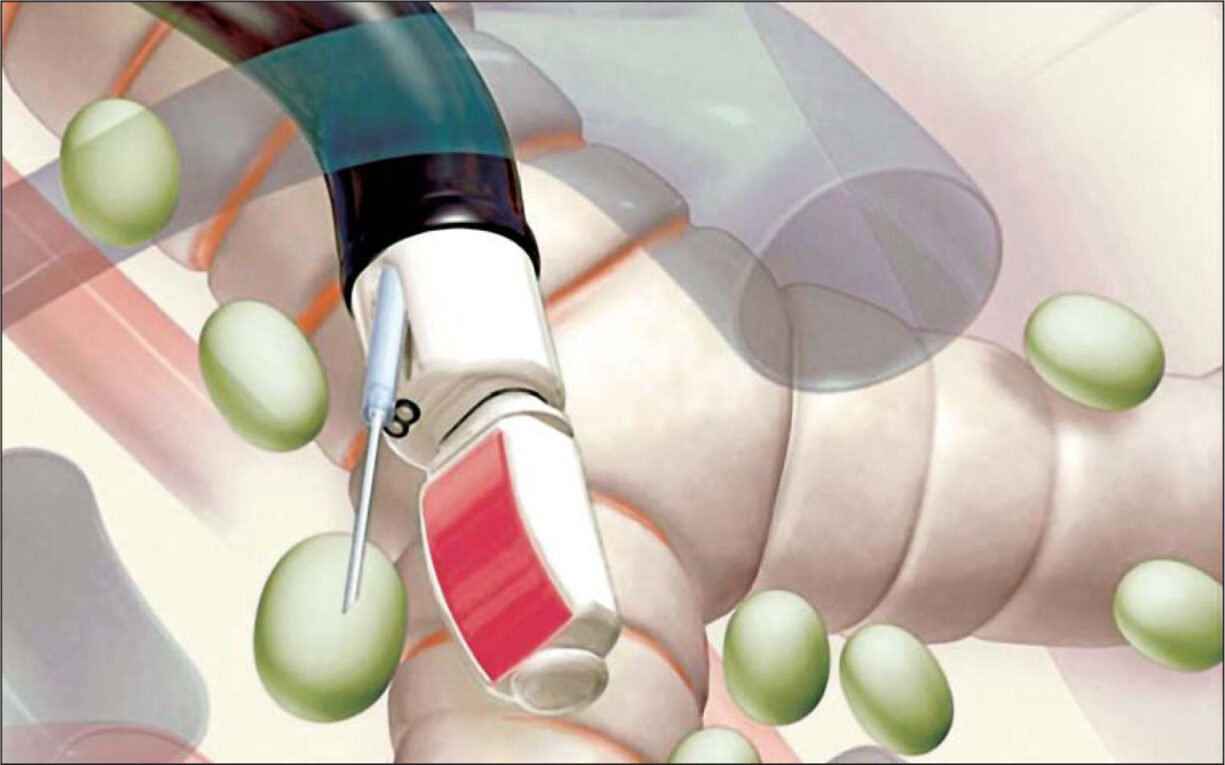
What is Endobronchial Ultrasound Biopsy?
Endobronchial ultrasound biopsy (EBUS) is a minimally invasive medical procedure that uses an ultrasound bronchoscope to view lymph nodes and tissues around the lungs and collect biopsy samples. The procedure allows doctors to get tissue samples from the lymph nodes and surrounding areas without open surgery. By examining the tissues under a microscope, doctors can diagnose conditions like lung cancer, sarcoidosis, and infections.
How is EBUS performed?
An EBUS procedure is typically performed in an outpatient setting under local anesthesia and conscious sedation. The patient is given medication through an IV to relax them and relieve any discomfort during the procedure. Then, a thin, flexible tube called a bronchoscope is passed through the mouth or nose into the lungs. The bronchoscope has an ultrasound probe at its tip that sends out high-frequency sound waves and uses the echoes to produce images of lymph nodes and tissues surrounding the airways in real-time.
Under ultrasound guidance, the doctor can precisely guide fine needle aspirates through the bronchoscope to collect samples from the targeted area for analysis. In some cases, a biopsy forceps tool may also be advanced down the working channel of the bronchoscope to collect tissue samples. The biopsy samples are then sent to a pathology lab, where a doctor examines the cells under a microscope to check for any abnormality or disease. The whole procedure usually takes 30-45 minutes to complete.
What areas can be sampled with EBUS?
Endobronchial Ultrasound Biopsy allows doctors to safely obtain samples from the mediastinal and hilar lymph nodes as well as masses that cannot be reached by standard bronchoscopy. Some of the key areas that can be biopsied using EBUS include:
– Mediastinal lymph nodes – Lymph nodes located in the middle compartment of the chest.
– Hilar lymph nodes – Lymph nodes located around the major airways (bronchi) within the lungs.
– Peribronchial tissues – Tissues surrounding the bronchi that may involve lung cancers.
– Lymph nodes above and below the diaphragm – Nodes above and below the major muscle separating the chest and abdomen.
– Lymph nodes along major blood vessels – Nodes along the vessels leading to and from the heart.
This allows for thorough staging of cancers to determine if they have spread from the lungs to other areas. EBUS helps provide targeted biopsies from specific lesions under real-time ultrasound imaging.
What are some advantages of EBUS?
Compared to other biopsy methods, EBUS has several important benefits:
– Minimal invasiveness – The procedure uses a thin, flexible tube so there is no need for open surgery. This results in less pain, scarring, and quicker recovery times for patients.
– High diagnostic accuracy – Studies have shown EBUS can detect more malignant lung lesions and metastatic lymph nodes compared to standard bronchoscopy alone. Its ultrasound guidance allows for precise biopsies.
– Assess distant lymph nodes – EBUS allows sampling of lymph nodes in the mediastinum that cannot be reached by other techniques like standard bronchoscopy or CT scans. This helps determine if cancer has spread.
– Real-time imaging – Ultrasound imaging provides real-time visualization of lymph nodes and other structures during biopsy, increasing safety and diagnostic yields.
– Outpatient procedure – EBUS is usually performed as an outpatient procedure under local anesthesia with mild sedation. Patients can often go home the same day.
– Combined with other modalities – EBUS can be combined with techniques like endoscopic ultrasound guided fine needle aspiration (EUS-FNA) for sampling both central and peripheral lung lesions through one procedure.
What are the risks of EBUS?
Overall, EBUS is generally a very safe procedure. However, as with any medical procedure that involves penetrating the body, there are some risks that can include:
– Bleeding – There is a very small risk of minor bleeding at the biopsy site that usually stops on its own. Severe bleeding is extremely rare.
– Infection – As with any invasive procedure, there is a slight risk of developing a local infection which can usually be treated with antibiotics.
– Pneumothorax – A small amount of air can escape into the space between the lung and chest wall, called a pneumothorax. This occurs in about 1-2% of cases but usually resolves on its own.
– Chest pain/discomfort – Soreness, pain, or discomfort in the chest is a potential side effect after the procedure but can be managed with over-the-counter pain medications.
– Mediastinal emphysema – Rarely, air can track into the mediastinal space, causing mediastinal emphysema. This usually resolves without treatment.
– Aspiration – There is a very small possibility of aspirating fluid, bacteria, or particles into the lungs during biopsy. Proper technique helps minimize this risk.
However, these potential risks are generally minor and far outweighed by the diagnostic benefits of obtaining tissue samples. EBUS continues to have a strong safety profile according to most published studies.
When is EBUS recommended?
Some common indications for an EBUS biopsy include:
– Evaluating suspicious lung lesions seen on imaging tests like CT scans that cannot be diagnosed by standard bronchoscopy alone. This helps determine if the mass is cancerous or not.
– Staging and sampling enlarged mediastinal lymph nodes in patients with known or suspected lung cancer. This helps determine if the cancer has spread beyond the lungs.
– Diagnosing sarcoidosis – a chronic inflammatory disease where enlarged lymph nodes are biopsied to look for non-caseating granulomas.
– Obtaining specimens from fungal infections or other granulomatous diseases of unknown origin.
– Sampling enlarged lymph nodes for diagnosis of tuberculosis or other infections.
– Guiding placement of fiducial markers in enlarged lymph nodes to aid in radiotherapy treatment planning for lung cancers.
In many institutions, EBUS is now considered the first-line test for evaluating mediastinal lymph node involvement in lung cancer since its high diagnostic accuracy. EBUS helps determine the appropriate treatment strategy.
Endobronchial ultrasound biopsy has emerged as an invaluable tool that has transformed the diagnosis and staging of lung cancers and other chest diseases. Its ability to safely obtain high-yield diagnostic samples from hard to reach lung structures under real-time imaging guidance provides crucial information. This helps physicians determine the most effective treatments while avoiding unnecessary invasive procedures. With benefits of precision, safety and comprehensive sampling, EBUS continues to grow in its use worldwide as the investigation of choice for pulmonary lesions and mediastinal lymphadenopathy.
*Note:
1. Source: Coherent Market Insights, Public sources, Desk research
2. We have leveraged AI tools to mine information and compile it

