
Chemiluminescence imaging is a powerful chemical technique that enables researchers to visualize biological processes and interactions with high sensitivity. By harnessing chemical reactions that produce light, this technique allows for detection and quantitative analysis without the need for external light sources. In this article, we will explore the principle behind chemiluminescence imaging, different applications that utilize this technique, and the significant impact it has made across various fields of biology and medicine.
The Mechanism of Chemiluminescence
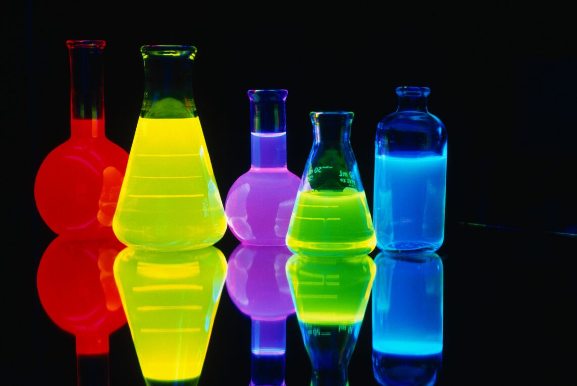
At the core of chemiluminescence imaging is a chemical reaction that emits light, known as chemiluminescence. This process occurs when an excited molecule or atom returns to its ground state and releases a photon of light. A common example is the oxidation of luciferin by luciferase in fireflies, emitting the familiar glow. In the laboratory, chemiluminescence reactions are triggered using stimulating reagents called luminol or lucigenin to produce a blue, green or yellow glow. By coupling these light-emitting chemical reactions to biological targets or processes, researchers can precisely visualize biological interactions and activities.
Applications in Molecular Biology
The high sensitivity of Chemiluminescence Imaging has made it a valuable technique for detection in molecular biology applications. Western blotting and ELISA assays commonly use chemiluminescent substrates such as enhanced chemiluminescence (ECL) reagents to detect proteins labeled with probes like antibodies. This allows visualization of proteins down to the femtogram level on membranes or in microtiter wells. Chemiluminescence is also used to quantify nucleic acids like DNA in techniques such as qPCR. By labeling PCR products or probes with chemiluminescent dyes, researchers can sensitively quantify gene expression levels during qPCR cycling. These applications demonstrate how chemiluminescence imaging enables precise quantification of molecular targets at extremely low levels.
Imaging Cellular Dynamics
Beyond detection on membranes or plates, chemiluminescence imaging has expanded visualization into living cells and tissues. Specialized probes have been developed that produce light upon activation by biological events inside cells. For example, calcium indicator dyes like calcium green emit brighter light as intracellular calcium levels rise. This allows real-time imaging of calcium signaling dynamics during processes like neuronal communication. Genetically encoded bioluminescent probes like luciferase have also advanced cellular imaging. Fusing luciferase to proteins of interest permits long-term tracking of protein localization, interactions and activity within intact tissues non-invasively. Overall, chemiluminescence imaging probes provide new insight into dynamic cellular and molecular processes in their native context.
Visualizing Disease Processes
The non-invasive detection capabilities of chemiluminescence in living systems have enabled new applications to study disease. Bioluminescence imaging using luciferase as a reporter gene is commonly employed to monitor cancer progression and responses to therapy in mouse models over time. Chemiluminescent reagents have also gained utility for in vivo bioactivation imaging of disease. For example, probiotic bacteria or viruses engineered to emit light upon activation by certain pathogens allow localization and tracking of infections in whole organisms. Oxidative stress beacons that luminesce in response to reactive oxygen species have potential for real-time imaging of tissue damage. Such tools advance our mechanistic understanding of disease while allowing non-invasive evaluation of new diagnostic and therapeutic approaches.
Chemiluminescence imaging principles continue to be combined creatively with new probe designs, delivering novel insights across biology and medicine. Areas like neuroscience and immunology have integrated chemiluminescent reporters to shed light on complex multi-cellular interactions in vitro and in vivo. Nanoscale chemiluminescence also shows promise for ultrasensitive detection on microfluidic platforms applicable to point-of-care diagnostics. Overall, the exquisite sensitivity and versatility of detecting light-emitting chemical reactions will ensure chemiluminescence imaging remains a powerful visualization technique driving major discoveries. As reporters and detection systems progress, its potential to illuminate life’s processes and advance human health will continue flourishing.
*Note:
1. Source: Coherent Market Insights, Public sources, Desk research
2. We have leveraged AI tools to mine information and compile it