
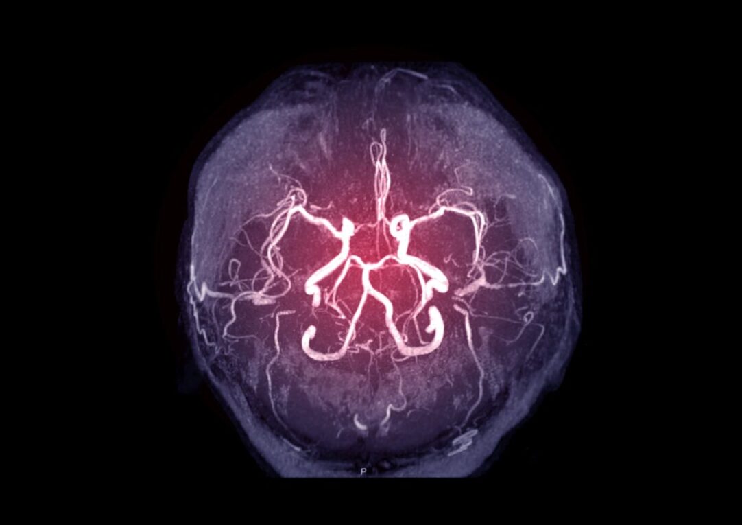
Cerebral angiography is a medical imaging technique used to visualize the arteries and veins in the brain and neck. It provides vital information for diagnosing and treating conditions that affect the blood vessels in the head and neck. Some of the common uses and indications for cerebral angiography are ischemic strokes, aneurysms, arteriovenous malformations (AVMs), tumors, and head trauma.
The Procedure
Cerebral angiography is performed in an angiography suite or radiology department. The patient is placed on the angiography table and connected to monitors to check vital signs like blood pressure and heart rate during the procedure. The radiologist first locates the femoral artery in the groin area and inserts a thin hollow plastic tube called a catheter. Guided by live x-ray imaging, the catheter is then inserted through the femoral artery and carefully maneuvered up into the arteries of the neck and finally into the blood vessels of the brain. Once the catheter has reached the arteries of interest, a special dye called contrast medium is injected through the catheter into the blood vessels. As the dye flows through the blood vessels, a series of x-ray images called angiograms are taken to map out the arteries and veins. The images clearly show up any abnormality, blockage, narrowing, tumors or other issues. After the procedure, pressure is applied to stop any bleeding and the catheter is withdrawn. The whole procedure usually takes between 30-90 minutes depending on the complexity.
What is Being Assessed?
Cerebral Angiography allows the visualization of even the smallest arteries and veins in the brain down to a few millimeters in size. This gives the diagnostic information needed to assess various cerebrovascular conditions:
Narrowing or blockages in arteries: Cerebral angiography can detect atherosclerotic plaques, thromboses or other causes of narrowing or obstruction in brain arteries. This helps diagnose ischemic stroke and identify areas at risk for future strokes.
Brain Aneurysms: Aneurysms appear as abnormal bulges or outpouchings in the artery wall. Cerebral angiography is one of the most accurate ways to detect, locate and determine the size of cerebral aneurysms.
AVMs: Arteriovenous malformations appear as a tangled cluster of abnormal blood vessels directly connecting arteries and veins without an intervening capillary bed. Cerebral angiography can diagnose these congenital vascular abnormalities.
Brain tumors: Some tumors like meningiomas and gliomas induce the growth of new blood vessels. Cerebral angiography helps show tumor-feeding blood vessels and assess the tumor margins.
Head trauma: In head injury cases, the angiogram can detect vascular injuries like tears, dissections and hematomas not visible on regular CT or MRI.
Potential Risks
Cerebral angiography is generally a low-risk procedure when performed by an experienced radiologist. Nonetheless, there are some potential risks associated with it:
Allergic reaction: A very small number of patients may have an allergic reaction to the iodine-based contrast dye used.
Bleeding: There is a small risk of bleeding from the groin puncture site. Serious bleeding is rare but possible due to the catheterization of arteries.
Infection: As with any invasive procedure, there is a slight risk of developing an infection at the catheter entry point. Proper sterile technique minimizes this.
Stroke: Rarely, pieces of damaged arterial plaque can break off during manipulation of the catheter and lodge in distant blood vessels causing a stroke. The risk is estimated at less than 0.1%.
Vasoconstriction/vasospasm: In some cases, the contrast dye may cause blood vessels to constrict excessively, which can transiently reduce blood flow. This is usually short-lived.
Nephrotoxicity: In patients with poor kidney function, the iodine contrast carries a risk of further worsening kidney function or failure. Precautions are taken in such individuals.
Overall, when performed by an experienced radiologist with proper case selection and informed consent, the benefits of cerebral angiography usually outweigh the minimal risks. The information obtained from cerebral angiography is invaluable for managing various cerebrovascular pathologies.
Cerebral Angiography in Clinical Practice
Let us now see some of the important clinical applications of cerebral angiography:
Diagnosis of Ischemic Strokes: Angiograms reveal thromboses, emboli, stenosis, and arterial dissections causing ischemic strokes. This helps initiate targeted treatment.
Detection and Classification of Brain Aneurysms: Cerebral angiography remains the gold standard investigation for detection and morphology assessment of aneurysms.
Pre-operative Evaluation of AVMs: The precise angioarchitecture of AVMs revealed helps decide treatment options of either surgery, embolization or radiosurgery.
Pre-operative Assessment of Brain Tumors: The vascular supply and relationships of brain tumors near major vessels are correctly evaluated before surgery.
Endovascular Procedures: Neurointerventional procedures like aneurysm coiling, carotid stenting, AVM embolization require cerebral angiography for guidance and procedure monitoring.
Follow-up after Surgery/Procedures: It helps assess the effectiveness of surgeries or endovascular treatments for cerebral vascular abnormalities.
cerebral angiography is an invaluable minimally invasive imaging tool that provides detailed anatomical information about the vessels supplying the brain and neck. It has revolutionized the diagnosis and treatment of various cerebrovascular disorders from ischemic strokes to vascular malformations. With experienced operators and careful case selection to balance benefits versus risks, cerebral angiography continues to be a mainstay investigation for cerebral vascular diseases. The high-quality angiographic images allow neurologists and neurosurgeons to accurately diagnose conditions and plan targeted management strategies. Cerebral angiography therefore remains an important component for multidisciplinary management of brain vascular disorders.
*Note:
1. Source: Coherent Market Insights, Public sources, Desk research
2. We have leveraged AI tools to mine information and compile it